|
|
|
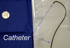
| Procedure DayStep-by-Step Details of Procedure Day How is UFE accomplished? Through a tiny skin nick at the groin we access the femoral artery with a small tube (catheter) that we manipulate into the arteries feeding the uterus.
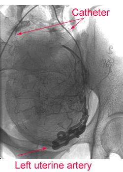
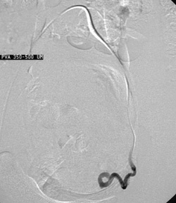
We perform an arteriogram, a roadmap of the arteries, by injecting an iodine-containing fluid while taking several digital x-rays. Here is an example of a digital arteriogram of the left uterine artery; the arteries are dark.
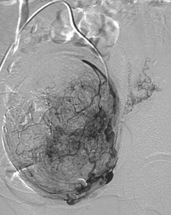
To see the blood vessels better, we subtract out the bones and background from the picture. This is called a "digital subtraction arteriogram" or DSA.
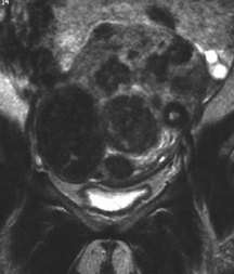
To better understand the relationship of this artery to the uterus, fibroids, and left ovary, I will superimpose this arteriogram on the MRI.
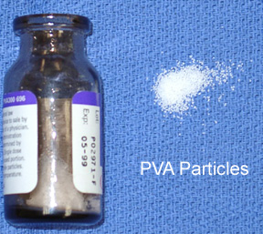
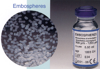
Then we inject a slurry of tiny particles that are carried downstream in the arteries to the uterus and fibroids. We use either particles of polyvinyl alcohol (PVA) or a relatively new type of agent called Embospheres. Blood flow to the fibroids is so much higher than the uterus that the vast majority of the particles are carried to the fibroids. These particles then block flow through the arteries. The image below shows how flow has decreased in the artery after UFE. Once the flow is adequately slow, the catheter is removed and we apply pressure by hand to the puncture site. Then we cover it with a band-aid: no stitches, no staples.
For more information, please contact me by e-mail or by phone 210.575.4343. Or visit the Society for Cardiovascular and Interventional Radiology's www site and follow the links about UFE.
 I subscribe to the
HONcode principles
I subscribe to the
HONcode principlesof the Health On the Net Foundation
This page was last updated on Tuesday, November 26, 2002 Comments about this site webmaster@drjohnthomas.com
|