|
|
|
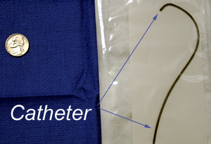
UFE is performed in the angiography suite. From the common femoral approach, we map out the arterial anatomy of the uterus, then use selective catheterization to gain safe purchase into the uterine arteries.
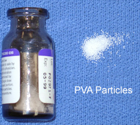
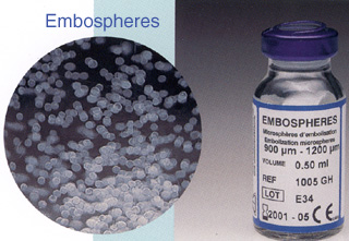
These are evaluated to ensure that embolization is safe (ex: absence of AV fistula or communication with bowel). Typically, a microcatheter is advanced coaxially deep into the uterine artery, past the cervicovaginal branch if possible. The artery is embolized with permanent particulate material. We use either 300 to 700 micron diameter polyvinyl alcohol (PVA) or 500-1200 micron diameter Embospheres.
Particles of this size occlude at the arteriolar level but not the capillary level. The abnormal, hypertrophied vessels supplying the fibroids will receive many more of these particles than will the normal uterine branches due to the increased flow.
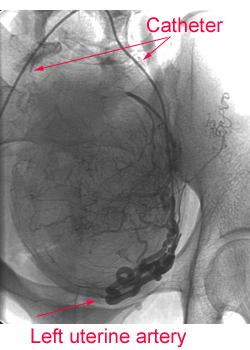
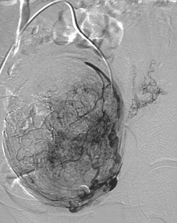
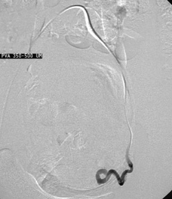
I usually embolize the left uterine artery first, then I manipulate the catheters into position to embolize the right uterine artery. The hypervascular fibroids, now devascularized, undergo fibrinoid necrosis. The uterus survives. Here are pre-embolization and post-embolization arteriograms of the left uterine artery. Pre-embolization Post-embolization
For more information, please contact me by e-mail or by phone 210.575.4343. Or visit the Society for Cardiovascular and Interventional Radiology's www site and follow the links about UFE.
 We subscribe to the
HONcode principles
We subscribe to the
HONcode principlesof the Health On the Net Foundation
This page was last updated onTuesday, November 26, 2002 Comments about this site webmaster@drjohnthomas.com
|